My routine.

Last update line to this post (7/13/12). The summary is that I got started on making the first piece of my model and found something new that I needed to include as a design principle.

As of 6/4/2012 this is an unfinished post! Since it is a creative work in progress on exactly how I turn brain information into a piece of art, I thought there might be preliminary interest before I was finished. It’s also nice having a record of the process for my own benefit 🙂

Most recent update

6/20 I Have Dye!

I’m almost done with the Legend. My next steps are to Finish the Legend, revisit the material briefly, and sketch the piece. I should be deliberate about this too. Even though the parts are easy, I want to do them right.

Note: My intentions are to update a couple of times and then make a final post.

6/5 update: Added the final decision on a trio of ganglia and the logic from source A1

6/7 update: Complication due to ambiguous structure, and doing figures right the first time. Below…

6/8 Finalized the method of diagramming the in-put/out-put logic! I think it’ MUCH cleaner and more understandable.

6/10 The weekend is a time for mentally resetting. This is valuable. I want to explain this someday 🙂 Essentially I need to add a new major element to the design so things got a bit slow while I was thinking. Now I have a solution!6/19

6/12 My apologies on the lack of updates. I have to order a black dye that I can use in the plastic so it might be a few days…

I’ll get some figures into this post. (…and yes I realize that I put beads into my figures twice…)

6/18: The dye will be here today hopefully. That would let me start tomorrow. This stuff has been designed in my head for over two weeks now and I’ve been going a bit nutty wanting to start. But the wait was worthwhile though because a legend page is a great idea. This one is turning out kind of neat because it’s like a lab meeting for you guys like the kind I used to always do during graduate school. A lot more fun than the real ones of course! (Unless the science is fun even when drier like it is for me 😉 )

6/19 I have no dye. Otherwise this will be the best map legend ever, or it will be horrible:)   I’m at 3049 words and really enjoying myself.

***************************************************************************************************************************************************

Now hopefully I can get a routine down to get this thing going in earnest! This will be the day to start good patterns and habits.(This will please you if OCD, maybe…)

Create a routine and just do one for now and as long as necessary. In fact that is what I am doing here. Blogging the experience of my first research attempt on my first model piece so that you can get an idea about the level of detail, and the thinking processes that I was engaging in while I was figuring out what I needed for every piece so that I can say the info is “GOOD”. It’s a model for the following work that I can refer to for anyone who is curious.

For every one of these brain processing centers I need the following information:

  1. The logical decision that it makes/role that it takes.
  2. Every known structure that it connects to.
  3. The logical nature of each of those connections (activation, inhibition, etc…).
  4. The number of connections (if possible, it might be important).
  5. The ultimate beginning and nature of the information that it carries/conveys. (For this and #6 it is to keep us in the “big picture both functionally, and experience-wise”)
  6. The ultimate end and nature of the information that it carries. (See #5 for context)
  7. Known Epigenetic regulation (for future use)
  8. What it looks like (might as well have some more context…).

For most readers 1-6 are of decreasing importance. 7 has to do with cutting edge things that are so new I can’t really do more than book mark it so I can build something into the model later when my brain spits out visual solution that is sensible in the information hierarchy. 8 will be useful to people in medicine or brain science. They will be more likely to have a mental link to the shape and it will be more interesting.

Of course I will add more as necessary, but that should be a good place to start. I have a system, I have a plan, what is the first piece!

#1 Spinal Trigeminal Nucleus

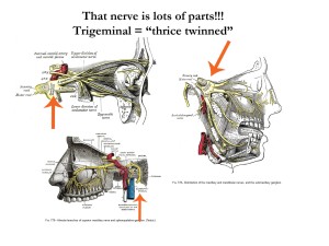
From Yahoo. Thank’s yahoo! (Trigeminal = “thrice twinned”. The freaking root is the “nerve”. Nerve is one of the most abused words in science. Its a linguistic whore…

Interesting context though. I almost feel like I’m in the room with Grey

Looks I have a more challenging piece for my first attempt. I like to Wikipedia first as a broad overview and it says,

The spinal trigeminal nucleus is a nucleus in the medulla that receives information about deep/crude touch, pain, and temperature from the ipsilateral face. The facial (cranial nerve 7), glossopharyngeal (CN9), and vagus nerves (CN10) also convey pain information from their areas to the spinal trigeminal nucleus.[1]. Thus the spinal trigeminal nucleus receives input from cranial nerves 5,7,9,10.

This nucleus projects to the ventral posterior medial nucleus in the dorsal thalamus.

That’s pretty bare for Wikipedia so either its pretty simple, or there is not much work with it yet. On top of that the source is gone. Would you trust that? The talk page just leads to a general medical portal, and the revisions just get simpler. This is when I start going to Pubmed, but later because this is a good example of what I keep running into when studying brain info:

Lack of specificity: Due to the long and complicated nature of brain research, this article uses historical Grey’s Anatomy images. This image is only useful when combined with other information because when Grey first did this work he numbered the cranial nerves I-XII, and their root ganglia I-XII. Since then they have discovered that these nuclei are more complicated than they thought because these nerves carry motor, sensory, place-in-space and other information from different places and for different purposes! So the Wikipedia page lists 12 cranial nerves, but 24 cranial nerve nuclei that translate and pass on the information. Annoying! The history has value, but that value is relative to the public’s utility. As long as the ultimate purpose of telling the history is honored in education (or fun), the information needs to be reorganized by everyone in biological sciences.

The Trigeminal Nerve, A TL;DR history.

It’s interesting that my first nuclei is actually mostly not about the body despite being at the bottom of the brain (or at least very low and central to the brain stem structure). That might say things about how human segments are arranged. In fact I think it suggests that the brain stem, should actually be thought of as the “Brain’s Spine”, combined with the information transfer of the “Body’s Spine” So the general information flow in the head looks like this…

…so far. In fact I think that a diagram of the information flow will be a necessary part of this project and I will draw one as I go. Fortunately Wikipedia even helps me here!

The brain stem is actually an interestingly complicated structure that literally routes information that impacts your entire body. It’s the most important information routing intersection in terms of overall structure at a “horror movie” or “that’s disturbingly interesting” level. So it’s actually a nice contrast that this structure has a complicated history when it comes to learning about it, and it being the first piece I make.

It was named as it looks, and the most obvious feature a centuries old anatomist would notice is that there are three huge nerves splaying from the cut zone. So yeah the old folks do the best with names that they can. The Trigeminal Nerve is interesting to me because the details of the information it handles shows us just how the “Human Worm Segments” of our head are arranged in gross detail.

Using Wikipedia on 6/4/2102, The nerves that feed this structure information mostly have to do with the face on the opposite side of the axis from the ganglion. But that information can be divided into two groups:

  1. Information about “…deep/crude touch, pain, and temperature from the ipsilateral face.”
  2. Physical routing of pain information from “The facial (cranial nerve 7), glossopharyngeal (CN9), and vagus nerves (CN10) also convey pain information from their areas to the spinal trigeminal nucleus.[1]. Thus the spinal trigeminal nucleus receives input from cranial nerves 5,7,9,10.” (We will see this link a lot. I will have to make a Link archive. I have to add proper citations to my routine anyway…)

The output is to the:

There’s that language again. Remember this old thing? I will need to make a new version with some more terms…

Ipsilateral means “same side”, Contralateral means “opposite side”. I think the entire reason that the brain does the side switching thing with information is to create a “Logical Contrast” so that it can tell a difference.

So we have the more “primal” type of pain, touch, and temperature from the face.

In addition we have unspecified pain information traffic for the face (facial nerve, “face/mouth nerve”), the mouth/throat(Glossopharyngeal nerve, “tongue-throat” nerve), and one of the most important nerves in your body, the Vagus nerve. Just pain information.

All that chaos above tells us that the ordered structure that we see in almost every human (there will always be exceptions) has rules, but we don’t understand them. So to properly understand the human body absolutely (if you are into that kind of thing) involves knowing the rules of the growth of the structures. That’s right, embryology.

Oh boy! A new toy! …and some old ones…

I know almost nothing about embryology, but the reason I am willing to play in an unfamiliar field is because I have enough experience with the fundamentals, and would have no problem internalizing a few new basic principles like the diffusion of chemicals in an embryo. (Sorry folks, it’s not a miracle. It’s only complex, which is why all the pain and suffering pops up too often.)

If you can see the results…

…you can get conceptual hold of any topic.

Embryology

and

Developmental Biology

Now I’m only going to include the bits relevant for us, but when development becomes important for functional understanding, I will bring it up. I guess in general I will need a couple of recent reviews on the structure/ganglia/nuclei mass just to make sure I have everything. I always try more than one kind of search so I don’t bias myself so I am trying (I already warned you there are cultural effects in the science and the medicine of the Brain/Mind).

A. [Spinal and trigeminal and nucleus], B. [Trigminal and Nerve]. 272, and 1680. That difference is due to the fact that one plugs into another, time, and because doctors don’t see a STN, they something that their intellectual ancestor called something like a “..special nucleus of gray substance…”, which is to say they don’t see a computer, they see meat.

For A, Sorted by date

  1. “The role of trigeminal interpolaris-caudalis transition zone in persistent orofacial pain.” is the most recent review that is really “meaty” and detailed. A
  2. “Neurochemical properties of the synapses in the pathways of orofacial nociceptive reflexes.” is a more “molecular/cellular/tissue angle. Not a review, but they will have background. It’s the kind of thing that I could actually explain if anyone was curious. I used to do something called “In-situ Hybridization” and it was fun! I made yeast cells glow red and blue and genetically modified the creature myself. Life can be legos sometimes. Although I have to assemble it myself with Word and screen grabs…
  3. “Anatomy of the Rostral Nucleus of the Solitary Tract.” A book chapter. I love these people. Not the same nucleus but also in the brainstem so will give context. More screen grabs…

For B

  1. This one is Meaty and fun if you have the stomach. A review investigating new scanning technology on a live patient! This is the benefit of broadening your search criteria on anything. Let your tastes guide you.
  2. “Non-Invasive Mapping of Human Trigeminal Brainstem Pathways” is the brainstem relevant, uh yeah… more Word and screen grabs though…
  3. If I need one after the fun stuff above…

I think that is a good start so Now I read and diagram out a mini logical structure of the region of the general Trigeminal nerve nuclei, out one heirarchy level from the the Spinal Trigeminal Nucleus. I have defined my artistic link to the brain…

6/5/2012

After reading “The Role of Trigeminal Interpolaris-Caudalis Transition Zone in Persistent Orofacial Pain” (A1 above) I have come to the conclusion that I must have a nod to actual structure in the model. But how to do this and not have it overwhelm the computation? I may just do it in metal wire that I can paint with a UV reactive substance so you can see the structure “light-up” over the computer bits…

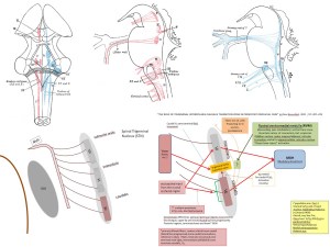
The rear/bottom part of the STN is the Vc (Nerve V, caudal nuclei) This does the pain and crude touch part of this nucleus. It will be Yellow plastic with Black and Red glitter. The logic of the connections that I could strip out of “The role of trigeminal interpolaris-caudalis transition zone in persistent orofacial pain.” is in this image. Looks like I’m building in interesting trio and then attaching the group!

6/7 Update

So where is that model piece you promised!

The reason this was done in a pseudo-“live blog” was that I knew complications would arise and that I would need adjust things. I am prone to internalizing bad habits. It take effort for me to get a bad routine out of my head and I love routines! So with something as long term and detailed as this I need to get it right early. So what is at issue?

  1. That really shitty diagram above
  2. ambiguous structures.

One is easy but it took me some time yesterday to think it through. I decided to take the Grey’s anatomy images and convert them to figures in Microsoft PowerPoint (you can do SO MUCH with MS office and Photoshop. Did you know you can copy/ paste from one to the other in many cases?).

That still needs some work but is MUCH better than the previous. Still too busy for most readers, but better for now.

Two is a little harder. When I read that first source and discovered this Vi/Vc transition zone, I discovered that it was a very important network hub for a lot of important processes, and it’s still being intensely studied. So while this model is really adaptable to having elements replaced, I need a way to present things like this transition zone. I am working on the second source above (“Neurochemical properties of the synapses in the pathways of orofacial nociceptive reflexes.”) and it includes even MORE extra info about this same transition zone! I hope to have that integrated by the end of the day, after I figure out how to present this hub of a transition zone. It’s more fun than frustrating, but when I say I am going to do something I need to be more specific to your guys…

6/8

Final method of showing the in-put/out-put logic! Three separate diagrams for each of inputs, outputs, and “unknown logic”

Next, and hopefully last before modeling something, how do I show these extremely important inter-ganglial transition regions? They are extremely important apparently! Look at all those outputs! I need a way to make them seem obvious when differentiated from the ganglia proper, but still related to the functional class…

6/10 Post one.

I think having a period where you just have fun and relax will one day be a constitutional right if we can define it. Chances are when I am done here we will discover that creativity as a process requires spacing and thinking. The more employers dump on employees, the more mental reset points will probably be necessary. This would probably be to ensure that the physical basis of practice and more importantly, internalization is achieved under the least stress. You can take that as a scientific hypothesis…

To make up for the weekend gap I am now adding a “Legend Page” where the design elements will be explained in specific terms, and the way the elements interact will also be explained so that you can understand how the picture becomes the data that create our experience. In general terms at least. Since science will always be adding to what we know about ourselves, this model is by design extremely easy to alter by nature. It’s an expression of the scientific method.

Here is a preview since it is still in progress. I hope I “code in” permanent elements today at the very least.

Colors and Sorting. Final decisions so I can start modeling ganglia.

I had a really cool post on philosophy and neuroscience planned, but I was too sick to brain that much so it became petty color day last week.

I’m starting this model out from the bottom where the spine becomes the brain stem. There really is no set place where “Brain” starts and “Spine” stops. I have a couple of places in mind, but the outline on the very bottom of the wire frame is based on spinal segment C2. So I might just start with everything neural from there.

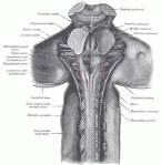
Left: Course of wires (nerve bundles) in the Brain Stem. Center: Anatomy of the nerves, muscles, and circulation of the upper neck and brain. Cerebellum removed. Right: Location of all Cranial nuclei I-XII.

The following is the organization that I am using to convey information on the nerves using color and pattern. The “Wires” of the body, the nerves are kind of complicated in science and medicine.

Doctors are unbelievably awesome people in terms of what they do, but training them has become a very intense process that is hard to change. So they all still learn the body by all the little complicated Latin names that have some meaning the tells you what the part looks like, and where it is. Because of that, The Original “Grey’s Anatomy” is still useful, and publicly available.

Right: Brain Stem with parts of Cerebellum removed, Rear (Posterior) view. Center: Cross-section of spinal cord showing Control (Motor, Red)) and Command (Somatosensory, Blue. Top is Rear/Dorsal, Bottom is Front/Ventral, and Supplemental Computer Control (Spinocerebullar, Blue also *sigh*), Right: Brain Stem from the Rear/Side (Poster Lateral)

But scientists who do all the mad scientist stuff that needs to be done to understand brains, talk in microscopic structures, and chemistry, and tissue, and genetics. So color and pattern is really the only thing I can use to translate that divide.

Wires

  • The wires going into the brain stem are the nerve bundles in the spine. These bundles represent outgoing command signals for movement, and incoming signals for data that we interpret as some of the classical five senses. These bundles are highly organized and the nature of the data is known to pretty detailed levels.
  • Here is a some general information on the nature of the white matter spinal nerve bundles. These are called “ascending tracts” or “descending tracts” which makes sense. You will also see “afferents” which is ascending, and “efferents” which is descending (link above).
  • You would feel it as “Movement” and “Touch/Pain/Hot/Cold”. To science it is Somatosensory data and Motor control.
  • This is a complicated model…

After staring at the information for a while I came up with the following set of information that I needed for every sense.

To the right is a screen grab from an Excel spread sheet where I am using to partially organize all of this craziness. These are all significant features that give me information that helps me to understand the data that these wires carry.

Commonly understood sense: This is not what you think. This is not Touch, Taste, Smell, Sight, and Hearing. This is the Somatosensory system, Gustation, Olfaction, the Visual System, and perception of sound. I’m afraid it’s much more complicated when you are an object instead of a subject. All that symbology…

Organ/system: Location: “sensor” substructure: This describes the physical nature of the minimal “thing” that detects the sensation after the simple receptor which would only bind a chemical, or move, or something. Examples include Muscle spindles that detect muscle stretch and let you unconsciously know where you are in space, or the Eye and it’s organized receptive fields.

“Receptor” ultimately responsible for “signal”: This is what we perceive from a single kind of receptor that detects something that we can interpret in experience. I got a lot of the ideas for whole list by looking at the receptors that we use to detect touch from here. What this describes are what is known in general terms about the different ways that we “feel” and these ways are defined at least partially by the receptor and the type of wire that carries the sensation. Examples of senses underlined as there were presented: muscle length and velocity, muscle tension, joint movement, joint angle, joint torque, touch flitter or movement, vibration, skin stretch, touch movement, fine touch, sharp pain or cool/cold, Dull aching pain or touch or warm…(always dot dot dot…)

Sensation common language: What you would call the feeling. I had those above.

Processing variations: This is where the real complexity starts. I got this idea from seeing how pain is processed and discovering the fact that there are at least three levels to how your body processes pain. These are called the Neospinothalamic tract, the Paleospinothalamic tract, and the Archiospinothalamic tract. Basically New, old, and ancient pain processing pathways. Think about it, do you react the same to a pinprick and a pool-cue through the abdomen? The “thalamic” part refers to the Thalamus which is a HUGE part of emotional processing and acts like a informational switchboard. One of my hypotheses is that these three divisions are mirrored in more than just pain perception. I believe that these represent three computational modes that we engage in and they have a huge impact on everything we do.

Body Region specific info: I am not ruling out the fact that there may be unknown information that body region can give me.

General Reflexes: Most of what you sense is carried out below the conscious level. Did you know you have multiple pathways controlling your movements and only one is “consciously controlled”? Many reflexes such as the stereotyped knee-tap don’t even need the brain at all except to let you know that it happened. I wonder if there are social reflexes…

Axon:Group:Diameter/Velocity: This refers to the behavior of the data as it travels along the wires in terms of speed and intensity. This matters A LOT. The next time you get a short stabbing pain pay attention. There is a noticeable distance between the time you feel the stab, the when you feel the throb (especially if you get a finger tip crushed). That is the difference between Aα and C fibers

Lower Body/Upper Body Routes: Just like peeking into the innards of an airplane, your wires are bundled too! 1′ refers to the first single cell in a series of connected wires. Some can go from your toe to your brain, one cell. There is usually a processing center at the site of connection and things you are consciously aware of tend to have very few stops. 2′ is the second wire that the first ware attaches (synapses) to. 3’… Reflex arcs might need information presented differently here since the paradigm changes a bit.

Face Routes: The same general principles but here things get complicated by the fact that humans are segmented like insects. It’s just that their segments are easier to see than ours. In general each human segment is represented by the bit where one nerve and vein/artery pair travel off of each side. So if you look at human segments this way you want to understand what is traveling in the Cranial Nerves and their ganglia.These have a very confusing course and they will be one of the later parts of the model.

If you were to stretch out a human by the segments and look at the function, we would be pretty weird. I’m sure that even more detail will come out later but for now…

  1. Smell
  2. Sight
  3. MoveEye/EyeRespond
  4. MoveEye
  5. FeelFace/Chew
  6. MoveEye
  7. ShowEmotion/Taste/Salivate
  8. Hear/SenseGravity/SenseMovement
  9. Taste/Salivate/Swallow/Speak
  10. RestAndDigest/Swallow/Speak/MoveTongue
  11. RestAndDigest/Swallow/Speak/MoveHeadAndNeck
  12. MoveTongue/Swallow/Speak

  • Side Note : You would be correct to think that I really, REALLY want to explore the evolutionary consequences of that series. Part of it involves understanding something called Hox genes, part is evolutionary biology, and part is developmental biology. After all if you look at a fly, or a Worm or two (Lots of cousins to choose from), you see really weird things. It’s thought segmentation evolved more than once. In fact the starfish is technically a bilateria, but somehow regained radial symmetry! That is a weirdness that I intend to pursue

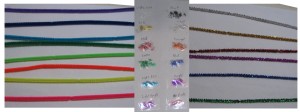
Representing Nerves: Spinal Wire Data. Pipe cleaners, glitter, and tape!

I have these pipe cleaners and glitter. Buying this with a Dremel tool got me really funny looks…

Options for showing differences in the same color of pipe cleaner include a strip of clear tape with glitter of one, or multiple colors depending on the information transmitted. I could also use a small piece of colored electrical tape or duct tape to add new layers of information that I might need. The nature of the connections that the wires make will be represented with colored paper clips (below). All of this can be changed easily if I discover a better way, but I have to start somewhere…

In the above Spinal cord cross section, I mentioned that there were Command (descending paths, or efferents, Red), and Control (ascending paths, or afferents, Blue) lines that represented the data that travels up and down your spine.

  • Side Note: some afferents and efferents are part of the same system, such as cerebellar  feed back loops that help with balance (The Cerebellum is pretty much the biggest regulatory computational region the brain has. That’s why it looks like a small brain. It’s big and old too…). They had to start somewhere so “This in/That out” was what they started with…

Motor

These command lines are in two categories.

The first are the voluntary, conscious pathways that you use to directly control your movements. I use White pipe cleaner because it tends to be culturally represented as “Good” in a neutral, universal way (I have ideas about this that do not involve race…). You are trying to move, it could be good or bad. The data that controls your conscious movement comes in three paths. The first two, the Anterior/Lateral Corticospinal tracts (Front/Side), control most of your movement. The Lateral path is the main path and controls arm and leg muscles for the opposite (ipsilateral) side of your body. The Anterior path also controls opposite arm and leg muscles, but for muscles located more centrally to the body. I am using Black/White tape to separate the two paths. The remaining voluntary path, the Rubrospinal tract, is an interesting path. It and the corticospinal tract can make up for the absence of the other so it seems to be some kind of extra level of movement control, but it is much smaller in humans than it is in other animals. I am marking that path with Red tape.

The second are the involuntary command lines that your body uses to make your movements correct, smooth, and precise. The Retuculospinal tract is responsible for other things as well, but this part is helping with balance and posture. Orange and Red Glitter are chosen because they are controlling you (from one perspective) and work together to do similar things. The Tectospinal tract is responsible for helping you to keep your head on a particular target. I thought the Light Blue glitter was good for vision. (Not to be confused with the Spinotectal tract, below). The Medial Longirudinal Fasciculus is another complicated tract. But a good part of it is devoted to similar roles as the TectoSpinal tract, controlling eye movements. It is Green glitter (also good for vision). Note that the poorer the knowledge of the function is, the worse the naming gets. Yeah, Tectospinal means Tectum-Spine in path. That’s it… Finally detection of gravity, motion and rotation and use of that to maintain posture is the job of the Vestibulospinal tract. I make that Purple because purple is what they use for royalty. A powerful force that can be good or bad…

Voluntary Motor

Involuntary Motor

Sensory

I have to apologize but the organization changes a bit here in the text because I have to adapt to the brain info and not the other way around.

Yellow pipe cleaner is the Fasciculus Gracilis or Cuneatus sensory nerves that carry “Place in Space” (proprioception), Deep Touch, Vibrational Touch, and Viscreal (organ) Pain) for the Lower Body, Trunk, and Upper Body respectively.They will simply be physically separated because they are easy to see.

Silver glitter on a Yellow pipe cleaner is “Place in Space”

Black glitter on a Yellow pipe cleaner is “Deep Touch”

Brown glitter on a Yellow pipe cleaner is “Vibrational Touch”

Yellow glitter on a Yellow pipe cleaner is “Viscreal Pain”

Blue pipe cleaner is the “Place in Space” proprioceptive information of the Dorsal, Ventral, and Rostral Spinocerebellar tracts. The nature of this information is still being investigated, but it certainly has to do with detecting limb and joint position. The Dorsal/Ventral pair work together and in different ways for the legs and trunk only. The Rostral tract does the same for the head and arms. Since other categories are likely as I look through the literature they will be Black and White as I think about what other information needs added. It’s the cutting edge in this section. Some sources report (I’ll flesh this out later) some touch and pressure as well for the dorsal tract so I will have to be careful or add other colors as I discover info.

Red pipe cleaner is the Lateral Spinothalamic tract because it carries pretty intense information: Pain and Temperature. Pain will be Red, Orange or Yellow glitter depending on the particulars (Sharp, throbbing and ???). The Anterior Spinothalamic tract will be Green in comparison because it carries Itch, and Crude Touch. Itch will be Red glitter, and Crude Touch will be Black.

The ascending Spinotectal tract is easily confused with the descending Tectospinal tract (above). So much so that Wikipedia is not as useful here, especially since this is still a mysterious tract. In fact their Anterolateral system page includes the wrong one! There are very few papers that look at the Spinotectal tract, but those that I can look at tend to think it has to do with detecting vibrations. That seems like a good use for the Purple pipe cleaners as any.

The Spino-olivary tract is yet another tweak involving propriception. This at least has a bit more research than the spinotectal, but Wikipedia is still not very useful.

Processing Centers (Ganglia)

It’ really interesting reading about brain organization. It’s like the brain has two mismatched “halves”, connected by a smaller brain. The top half they call the Cerebrum, the bottom half they call the Midbrain, and the small brain is the Cerebellum. If I were to assign identities to these parts I would call the Cerebrum “Consciousness and You”, the Midbrain “Why I’m doing this/How I feel about this/This is how you are doing/This is what you need to do” and the Cerebellum is “The Puppeteer”.

Cortex

There is a weird transition in structure from the Midbrain to the Cerebrum. The Cerebrum is mostly made out of Cortex, which I still need to decide how to represent. It’s basically layers of cells that you can even see. Somehow (they are still working this out and I need to read more) this region processes your conscious experience in columns that are also communicating with the regions next to them, and other regions of cortex by way of the Basal Ganglia.

The Cortex is a layered structure where most of the memory storage somehow involves connections between layers. “A” in this image is in the middle of the cortical layers. While this is not human cortex, the more animals that have a structure, the more basic it is to our common experience, the more general conclusions you can draw. The circled area above is the Hippocampus which is involved in long term memory formation and the meanings of other abbreviations can be looked up here.

Midbrain

The Midbrain is mostly nerve bundles and  Ganglia which act like little processing centers that make logical connections, and logical decisions. (There is supposed to be a little cortex in the Midbrain and eventually I will find that info. The kidney has adrenal cortex…).

Here is the Basal Ganglia which is particularly relevant to me because it is involved in both ADHD and Tourette Syndrome. You don’t need to look at them but I thought more examples of ganglia would be nice.  (A Couple of sources for this image)

Now that image is not quite done, but I just wanted to start it for now because I am trying to work on the ADHD and TS posts too. The take home message from the links is that the Basal Ganglia is/are part of how your brain prevents you from making mistakes, and it/they are constantly cycling though different connections in your cortex as you are consciously interacting with the world. It has a direct (activating) and indirect (inhibitory) path that is constantly making, and breaking connections between cortical regions. Guess which one I have problems with 🙂

Cerebellum

The Cerebellum is a structure that helps you to learn to do things, mostly motor things, and makes sure that you do things right like an ever present teacher that is holding your hand while showing you how to color without going outside the lines. There is a lot of evidence that is operates outside of the motor system too.

The only other feature worth mentioning is that there is a third “structure” in the brain that is the oldest, and least organized. It is called the Reticular Formation. It seems to be a looser collection of networks and ganglia that handle unconscious and very ancient tasks like balance. posture, sleep/wake cycles, cardiovascular control, respiratory control, pain, and habituation (the ability to learn to ignore things). This will be modeled, but will be challenging.

I first started by looking at anatomy in recent review articles like this one about the Brain Stem, and the Wikipedia entry. That was really important to get a general feel for the field and how it is described and discussed. But when I found this Anatomy text available for free (above image), that had to be how I started making the ganglia models.

This has 29 individual ascending sections of a human brain stem. This not only gives me each little tiny processing center in the brain stem (up until 2004), it gives me the nerves, and cells that have to do with neurotransmitters, and more that I can use for info later. In the previous post I pointed out how when I shaded these ganglia by functional class, the organization started to really show up well!

So right now I am entering all the names of the brain stem ganglia into a spreadsheet and looking up functions and links to papers so I can connect them in the model. I don’t know if I am unrealistically ambitious but I’m going to try damn it.

The list has over 300 items that I still need to categorize, but between that, Grey’s Anatomy, and the Wikipedia entry on the cranial nerves I am going to start with:

  1. Spinal trigeminal nucleus (Vs)
  2. Spinal accessory nucleus (11, or XI))
  3. Nucleus ambiguus (11, or XI)
  4. Solitary nucleus (Connected to IX and X according to Grey)
  5. Dorsal motor vagal nucleus (X)
  6. Central Cervical Nucleus
  7. medial pericuneate nucleus
  8. Medial Reticular Nucleus of the Medulla (and internal arculate fibers?)
  9. Inferior salivary nucleus IX
  10. Hypoglossal nucleus XII

Why? They are central, and low so would be the best place to start pragmatically. I’ll try to say something about them as I make them and display progress here. The stuff in quotations are things I’m still thinking about.

Representing Ganglia: Dyed InstaMorph plastic embedded with paperclips, dye and glitter! Thumb tacks used too!

I need a material that is fast and easy to use, modify, and place. This seems to work just fine Colors will be chosen similarly to the wires above. To represent a logical decision that is made by the wire/ganglia connection I will attach the pipe cleaner to the ganglia with a colored paperclip that will indicate the kind of connection. Red for inhibitory, Green for excititory.(Off or ON)

An adjacent thumbtack (actually I may changes this to a colored bead on the pipe cleaner) will indicate the neurotransmitter that is used in the signal transfer.

I will find another solution (like the bead) to represent any details having to do with the signal pattern of the firing, or the logical consequences of such (http://en.wikipedia.org/wiki/Neural_coding) (Example: spiking interneurons).

After the primary sensory wires meet the first data transfer centers, who knows what I will need to do to properly represent the way the information is transformed? This could get really interesting…

I’m just starting to realize how crazy this could get, but it feels possible…

Pant, pant, pant…


Why is brain science so difficult to understand anyway?

In which I take a step back…

First let me say that there are good reasons for the names and terms that need to be changed. When they were first bestowed they did not know what the parts did in the brain. Also keeping a name the same (no matter how silly) can make it easier to do science between folks from different countries. But that usefulness loses it’s relative value once the function of a part of the brain becomes better defined. At some point tradition has to give way to reality and

the name should be changed to something that represents it’s function so that non-scientists can use that information as soon as possible. Especially with something as potentially useful as brain science.

But here is where I can almost understand why there might be a genetic component to conspiracy theories. It might look like I’m seeing hierarchies everywhere, but it’s not crazy if it’s true and I can demonstrate it. A bunch of old sciences are joining together into something new and that new thing has to be useful to people. Brain science needs to do renaming and in a real way the organization of the colors in the model are a process for doing that.

Science and Naming

Science is a constantly evolving discipline because it is a process, and the things that come out of that process can change the outside appearance of the whole enterprise. Part of that is due to the fact that science sometimes gets things wrong and better information is found later.  But what is “wrong” is usually the best that they could do at the time. I don’t stick my nose up at alchemy because it was a hypothesis, and chemistry was it’s competitor. Chemistry won, for the most part (I suppose nuclear chemistry is close enough to alchemy in some respects ;)…) As an example the rhetorically abused “paradigm change” that occasionally does occur, such as the eventual acceptance of plate tectonics.

Here is a good story about Alfred Wegener and how he changed Geology.

From Wikipedia’s Continental Drift article.

But another part of that is also because multiple fields of study discover that they are really parts of the same thing and it can take a while to get everything worked into a whole with language that everyone uses. This happened with DNA. At first they knew nucleic acid existed, and that there was something in the cell that stored the genetic information. Eventually the right experiment was done and the two fields, DNA and genetics, were merged.

Here is Wikipedia’s description of the Hershey-Chase experiment which is the one they show us in Molecular Biology to tell the history of how they knew that DNA was where our genes were.

Martha Chase and Alfred Hershey. From The Cold Spring Harbor Laboratory DNA Learning Center.

Brain science is a lot like the second one except that these fields knew that they were related from the start. It’s just that the brain had so many dimensions to how it functioned that it’s study started in many different places at different times.

  • Molecular biology: The study of the nature and structure of the chemical networks inside of cells, focusing on the molecules that do the chemistry. Biochemistry (think chemistry specific to biology) is a big part of this field.
  • Cell biology: The study of the behavior of cells and the interactions between cells.
  • Neurobiology: The study of the cells of the nervous system and their interactions. This roughly blends into,
  • Anatomy of the brain, psychology, sociology, more!

The reason all the names and interactions suck to learn is that when these things were first being investigated, the scientists often times did not know what they were dealing with so they had to have something, anything to create differences that they could use to sort and study parts.

It started with anatomy.

Here is a free online version of Grey’s Anatomy (the book, not the comedy). Back then they had very little idea what this stuff was all about. There were some items known in general terms from specific brain injuries, but the structures were mostly all named according to what they looked like, in Latin, or French, or…

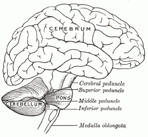
Cerebellum (little brain), medulla oblongata (middle body), median fissure (middle division). It can be a little intimidating.

Then there was Cell Biology and Neurobiology.

Cells had the same problem with naming. You have lots of names having to do with names, shapes, and now electrical properties as well. This was not just for the nerve cells either, your brain has more non-nerve cells in it than nerve cells believe it or not. The general name for those cells is Neuroglia, nerve glue! I’m sure at the time it was fine, but for everyone else? Here is a small selection;

Neurons: Pyramidial cell, Purkinje cells, uni- bi and multipolar cells…

Glia: Microglia, Schwann cells, astrocytes, satellite cells

On top of that now the cells are also being divided by electrical activity. This overview of the somatosensory system (touch/pain sensing system) shows cells being named by the speed of the impulse, the voltage peak (literally detected by micro voltmeter)

Now admittedly this last bit is far more useful because these features do give me information that can be used in organization and study. But I maintain that it is useless to regular folks who have a relative with a problem, or their own problem and a life to deal with.

Then there was modern brain science, and more crazy naming.

Here is where my issues with naming and science start to become unfair, which is why it’s a process and not a rule. If the role of a part of the brain is unambiguously known, or known well enough that it’s primary function is known, that is when the name should be changed. But when you are sitting on the cutting edge things are still too fuzzy for that. Here are some examples of things that can’t really be renamed that I will have to take into account somehow;

  • Structures/cells/molecules with still unknown functions and/or relationships.
  • Whole fields that are not yet known well enough to integrate into brain science.

Unknowns

They name these things whatever they have to to understand them. I will have to have a way of making “function unknown” visually obvious in a way that does not detract from visually appreciating what is known. I will probably have a color, or obvious texture, or similar reserved for that purpose. I’m hoping to make some basic decisions this week.

How carefully do I need to do this? Here is an image from one of my sources that I found freely available on the web. This is a section of your brain stem (there are 25 pages of these).

From “Organization of Human Brain Stem Nuclei” by Yuri Koutcherov, Xu-Feng Huang, Glenda Halliday, and George Paxinos of The Prince of Wales Medical Research Institute The University of New South Wales. Brain nuclei are literally small brains that have jobs in processing information for your body. The are information nodes in a net work, and they are sensible and understandable!

This is the most fascinating part of the brain to me because what I am most interested in are the very oldest things in our ancestors. If I had managed to get my PhD I wanted to study Origin of Life issues. This is close too!

The brain stem is the most ancient structure evolutionarily speaking. There is a rough progression from oldest to youngest as you move from the bottom of the brain stem through to the forehead. What that means is that if you find that you share something with a creature that is REALLY distantly related to you and it’s in the brain stem, you probably have something really mentally universal and fundamentally important. This is where the model starts.

The brain stem is the origin of ME, and YOU in that big grand ultimate sense that we mean when we want eternal life, the propagation of the self and our memories and experiences into continued existance. This is where Brains began because this is where an ancestor that was a worm-like thing first made* something “different” at the front in terms of how information flowed in it’s body. This is like a worms brain. This is;

  • Vomiting, Sneezing, Coughing (Generalized “Get the toxin out!”)
  • Breathing
  • Heart Beating
  • Sleeping (It’s that old…)

Go back much farther and you lose things that are bilateral. Yes, not long after a tube shaped body appeared evolution took advantage of cells that communicate in a way that made the “self” possible.

To start organizing it I printed out all these pages and started coloring things with colored pencils by category of function as best as I could. No particular color but I toyed with making certain some colors certain categories.

Just look at that organization! Sometimes I sympathize with Creationists. If you don’t know how to understand Embryology it can look deliberate. But in the end it’s still just the Arrogance of Ignorance…

I just want to start making hypotheses about why it’s organized the way that it is and what that says about our ancestors!

The book chapter lists these categories. In the parentheses afterward I will try to convey what they mean to you in a personal sense. The colors are matched to the colored pencil colors as closely as I could manage and the regions with black shading are the white matter (wires) and areas unshaded with black are the nuclei/ganglia (information processing centers). This is all human specific information and I will not include animal information until I get to the model.

  • Autonomic Regulatory Centers
  • Reticular Formation
    • The Brainstem’s brainstem. Very old parts. (Maintaining tone, balance, and posture–especially during body movements. Sends eye and ear signals to the cerebellum so that the cerebellum can integrate visual, auditory, and vestibular (balance) stimuli in motor coordination. Gaze centers, which enable the eyes to track and fixate objects, and central pattern generators, (programmed muscle movements) which produce rhythmic signals to the muscles of breathing and swallowing. Cardiovascular control.  Pain modulation.  Sleep and alertness (all or nothing conspicuousness?). Habituation – This is a process in which the brain learns to ignore repetitive, meaningless stimuli while remaining sensitive to others.
  • Tegmental Nuclei
    • Cognition, motivation, drug addiction, intense emotions relating to love. May be involved in modulating sustained attention or in mediating alerting responses, and also in the generation of REM sleep. Head direction.  Arousal, attention, learning, reward, and voluntary limb movements and locomotion.
  • Locus coeruleus involved with physiological responses to stress and panic.

I think you get the point. See how the language sucks. But there is so much information there that could be more helpful if it was better presented for the public.

They know TONS about how these bits connect together, what the bits do, and lots of the logic (computational rules like “On”, “Off”, more…) between them. If you are a philosopher and you are not reading neurobiology at whatever level you can, you are being professionally negligent. But it’s only partially your fault, hence this effort.

These networks and how they differ on an individual basis is what I hear used as the term neurodiversity in public. Ignore it at your peril. We are a spectrum, not black and white. Mental illness is the definition of the extreme. I know that some of the science readers here might get a bit uncomfortable with the term because there are a lot of bad ideas associated with some folks who use the term. But it is accurate and that is what I care about.

Stuff that is new in a BIG way

There are two that I can think of: RNA interference and epigenetics (I’m preparing a cool post about that, eventually). RNA interference is basically when RNA plays a role in shutting off gene expression instead of being a messenger molecule. Epigeneitcs is something that I will have to do a post on all by itself at some point. This is when a trait gets inherited for a limited number of generations, or gets inherited with no actual change in the DNA. What gets inherited are little “marks” on the DNA that change the expression. While I have not yet explored RNA interference and the brain yet, epigenetics is VERY involved in brain function. In fact I’m of the opinion that this will be next decade’s controversial research subject due to it’s implications.

So there is a bit more on the level of detail that I want to take this model. Right now I am entering nuclei into a spread sheet and adding relevant details about connections, role (human and animal studies), neurotransmitters, everything.

Next post. Something different. My weird relationship with philosophy.